[creative-diagnostics] Coronavirus Antigens
| 작성자 | 관리자 | 작성일 | 2020-02-21 |
| 조회수 | 4,383회 | 댓글 | 0 |
본문
바이오스토리에서"creative-diagnostics" 직수입 합니다.
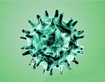
Coronavirus Antigens Browse All Coronavirus Antigens Coronaviruses belongs to one of two subfamilies: Coronavirinae and Torovirinae, are a family of enveloped, single-stranded, positive-strand RNA viruses classified within the family Coronaviridae, in the order Nidovirales. The name “coronavirus,” is derived from the “corona”-like or crown-like morphology observed for these viruses in the electron microscope. There are four proteins that contribute to the overall structure of all coronaviruses: the spike (S), envelope (E), membrane (M) and nucleocapsid (N) The type I glycoprotein, the spike (S) that forms the peplomers on the virion surface, giving the virus its corona- or crown-like morphology; the membrane (M) protein, a protein that spans the membrane three times and has a short N-terminal ectodomain and a cytoplasmic tail; and small membrane protein (E), a short ectodomain, a transmembrane domain, and a cytoplasmic tail. The genome RNA of Coronaviruses is complexed with the basic nucleocapsid (N) protein to form a helical capsid found within the viral membrane.
Fig. 1 Model of Coronavirus Coronavirus’ replication begins with the virus entering into the cytoplasm of cells, once the virus gets into the cells and then it releases its genetic substance into cytoplasm. The Coronavirus genome has a 5’ methylated cap and a 3’polyadenylated tail. This allows the RNA to attach to ribosomes for translation. With the help of replicase encoded in its genome which allows viral genome the RNA to be transcribed into new RNA copies using the host cell's staff. The replicase is the first protein to be made; once the gene encoding the replicase is translated, the translation is stopped by a stop codon.
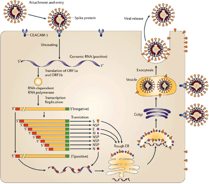
Fig. 2 Signal Pathway of Coronavirus’ Replication S/HE protein locating at the surface of Coronavirus binds cell through receptors on surface of cell. After binding viruses get into the host cell by fusion of viral and cell membrane or by receptor mediated endocytosis. Because coronaviruses have a single positive stranded RNA genome, they can directly produce their proteins and new genomes in the cytoplasm. At first, viruses synthesize its RNA polymerase which only recognizes and produces the viral RNA. Subsequently, negative strand serves as template to transcribe smaller subgenomic positive RNAs. The whole process of replication of Coronavirus in host cell shows in figure 2. Recombinant COVID-19 Nucleocapsid Protein (29-213 aa) [His] (DAGS029) Nature: Recombinant Expression System: E.coli Tag/Conjugate: His Application: ELISA, LFIA, WB
Recombinant COVID-19 Nucleocapsid Protein (59-213 aa) [His] (DAGS059) Nature: Recombinant Expression System: E.coli Tag/Conjugate: His Application: ELISA, LFIA, WB
Recombinant COVID-19 Spike Protein Receptor Binding Domain [Fc] (DAGC089) Nature: Recombinant Expression System: Human Cells Tag/Conjugate: Fc Application: SDS-PAGE
Recombinant COVID-19 Spike Protein 1 [His] (DAGC091) Nature: Recombinant Expression System: Human Cells Tag/Conjugate: His Application: SDS-PAGE
Recombinant COVID-19 Spike Protein 1 [human Fc] (DAGC092) Nature: Recombinant Expression System: Human Cells Tag/Conjugate: Fc Application: SDS-PAGE
Recombinant COVID-19 Spike Protein 1 [mouse Fc] (DAGC093) Nature: Recombinant Expression System: Human Cells Tag/Conjugate: Fc Application: SDS-PAGE
Recombinant COVID-19 Nucleocapsid Protein (Full Length) [His] (DAGC094) Nature: Recombinant Expression System: E.coli Tag/Conjugate: His Application: ELISA, LFIA, WB
바이오스토리에서"creative-diagnostics" 직수입 합니다. |